![]()
Thanks to a spectroscopy technique originally developed for materials science, water is becoming an original way of monitoring the biological response of cells to a specific stress, such as exposure to a photosensitive therapeutic agent.
Laure Gibot, Anne-Françoise Mingotaud and Patricia Vicendo from the IDeAS team, in collaboration with other scientists from the Laboratoire d'optique et biosciences de l’École polytechnique and the Centre de microscopie électronique appliquée à la biologie, have just demonstrated it, in a study published in the journal Advanced Science.
Terahertz (THz) spectroscopy is a widely used physical technique for characterising materials using electromagnetic waves. In a frequency range from a few hundred GigaHz to a few THz, it is accompanied by a strong water response that opens up potential applications for biological studies of water-rich tissues and cells. Unlike other imaging techniques, the radiation used in THz spectroscopy to probe the sample is safe for biological tissues because it does not physically interact with the material.
Thanks to this spectroscopy, this interdisciplinary consortium of French physicists, chemists and biologists has recently visualised and quantified the early phenomena occurring at the cell membrane level, following anticancer treatment by photodynamic therapy (PDT). This treatment, which is still not widely applied in France, uses photosensitive molecules. In the absence of light excitation, these molecules are non-toxic and inactive. However, following light activation and in the presence of oxygen, they locally generate oxidative stress which can lead to cell death in the case of anti-cancer medical applications.
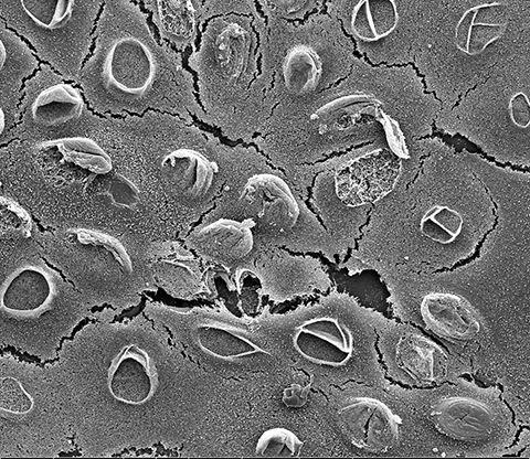
The scientists carried out these physical measurements using THz spectroscopy, in parallel with classical biological experiments in the laboratory. The effect of photodynamic therapy on cancer cells was thus demonstrated. The results obtained indicate a real added value of THz spectroscopy for monitoring the treatment compared to the usual biological approaches. Firstly, it allows them to observe the evolution of cellular responses from the start of the light irradiation, by monitoring the intracellular water content, in real time, and over several hours. By monitoring the number of defects (holes) created on the cancer cell membrane, it also allows a quantitative estimation of the damage caused by local oxidative stress during photodynamic therapy. THz spectroscopy is therefore a promising technique that complements classical and established biological studies and techniques.
Therefore, these results open up new perspectives in the understanding of biological phenomena inducing a modulation of the water content of the cell, as is the case in anti-cancer treatment by photodynamic therapy.
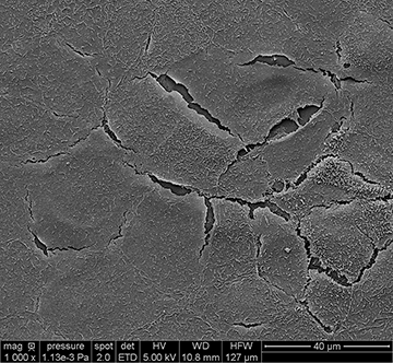
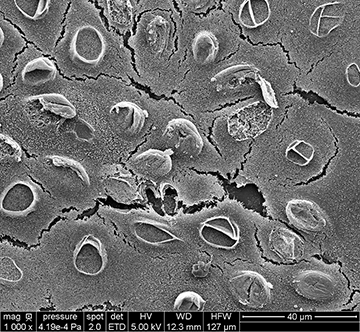
Observation by scanning electron microscopy of untreated cancer cells (control, on the left)
and treated ones with a photosensitive drug (PDT, on the right).
© Rachel Brival – IMRCP/CMEAB
![]()